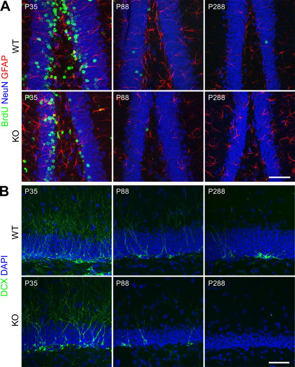
Figure 7.
Illustration of neurogenesis in the dentate gyrus of cD2KO and WT litters.(A) Sections of brains aged P35, P88 or P288 were stained against BrdU (green), NeuN (blue) and GFAP (red). Irrespective of genotype, the majority of BrdU-positive cells co-labeled with NeuN indicating that neurogenesis takes place also in the DG of cD2KO mice, albeit at a much lower rate. (B) Representative DCX-labeling (green) at P35, P88 or P288. CD2KO mice appear to have less DCX-positive cells than WT litters. Sections were counterstained with DAPI. The images in (A) and (B) are merges of multiple confocal planes (for NeuN, GFAP, BrdU: 4–5 planes spanning a z-dimension of approximately 4.8 to 6.4 μm; for DCX: 3–4 planes spanning a z-dimension of approximately 3.6 to 4.8 μm). Scale bars: 50 μm.

