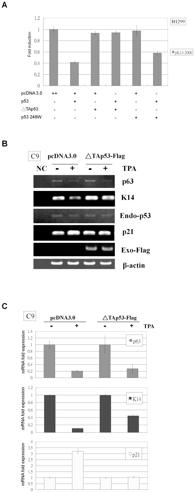
Figure 4. ΔTAp53 relieves TPA-induced K14 suppression in C9 cells.
(A) H1299 cells were co-transfected in a three-vector reaction with the pK14-2000 promoter vector, pcDNA 3.0 or wild-type p53 expression vector, and pcDNA 3.0, p53 248 W, or ΔTAp53 expression vector. The levels of luciferase promoter activity were normalized to the pcDNA 3.0, which was set to 1. Wild-type p53 repressed pK14-2000 promoter activity, but this effect was completely reversed when ΔTAp53 was co-transfected or partially reversed when p53 248 W was co-transfected. (B) C9 cells were transfected with pcDNA 3.0 or ΔTAp53-Flag and then were treated with DMSO or TPA. Following treatment, total RNA was isolated from each sample for RT-PCR analysis. p63 mRNA was down-regulated by TPA treatment, but endogenous p53 Mrna levels did not change after TPA treatment. K14 and p21 mRNA expression were down- and up-regulated by TPA, respectively, but these effects were diminished in ΔTAp53-Flag transfected cells.Anti-Flag antibody was used to assess exogenous ΔTAp53-Flag expression, and anti-β-actin antibody was used as a loading control. “NC” denotes the non-cDNA PCR control. (C) Total RNA was prepared from the same treatment conditions as in Figure 4B and was subjected to real-time RT-PCR analysis. p63 was down-regulated by TPA treatment in pcDNA 3.0- and ΔTAp53-Flag-transfected cells. K14 was down-regulated by TPA treatment, but this effect was partially reversed in ΔTAp53-Flag-transfected cells. p21 was up-regulated by TPA treatment, but this effect was fully reversed in ΔTAp53-Flag-transfected cells.

