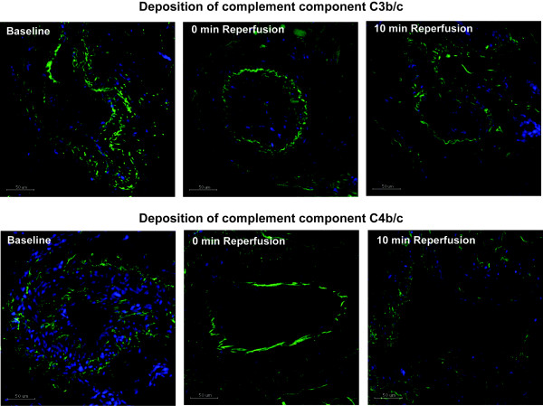
Figure 5.
Immunofluorescence staining for C3b/c and C4b/c. Tissue samples from baseline, end ischemia and 10 min reperfusion were analyzed by Immunofluorescence for deposition of the complement components C3b/c and C4b/c. Pictures are representative for the time points and no significant differences were found by Repeated Measures ANOVA, n = 10. Green = C3b/c / C4b/c, blue = DAPI staining of nuclei. Scale bars = 50 μm.

