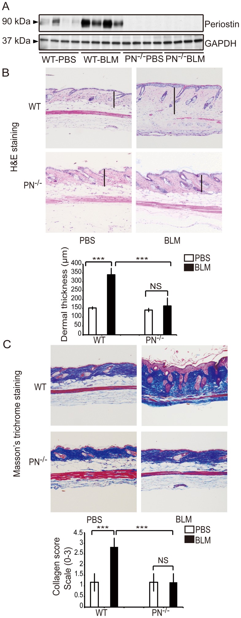
Figure 2. Periostin gene knockout (PN−/−) mice are resistant to BLM-induced cutaneous sclerosis as assessed by dermal thickness and collagen deposition.
A, Western blotting analysis for periostin in skin extracts from WT and PN−/− mice, which were treated with BLM or PBS. B, H&E staining of skin samples from WT and PN−/− mice (original magnification, ×100). Dermal thickness is shown as the black bar in the lower panel and was measured as described in the Materials and Methods. C, Masson’s trichrome staining of skin samples from WT and PN−/− mice (original magnification, ×100). Collagen fibers were stained blue. Collagen deposition was scored on a scale of 0–3 as described in the Materials and Methods and is shown in the lower panel. For all assays, 10 mice from each group were analyzed. Values in B and C are shown as the mean ± SD. NS, no significance; ***, p<0.01.

