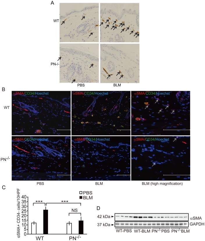
Figure 4. Periostin is required for dermal myofibroblast development in BLM-treated mice in vivo.
A, Representative skin sections from WT and PN−/− mice, stained by immunohistochemistry with anti-α-SMA antibody (original magnification, ×400). α-SMA-positive myofibroblasts are indicated by arrows. B, Representative skin sections from WT and PN−/− mice, doubly stained by immunofluorescence for anti-α-SMA (red) and anti-CD34 (green). α-SMA+ CD34− spindle-shaped myofibroblasts are indicated by arrows. Scale bar = 100 µm. Nucleic staining: Hoechst 33342 (blue). C, The number of myofibroblasts per 10 hyper power microscopic fields is shown in the histogram. D, Western blotting analysis of protein extracted from WT and PN−/− mice skin tissues. For all assays, 10 mice from each group were analyzed. Values in C are shown as the mean ± SD. NS, no significance; ***, p<0.01.

