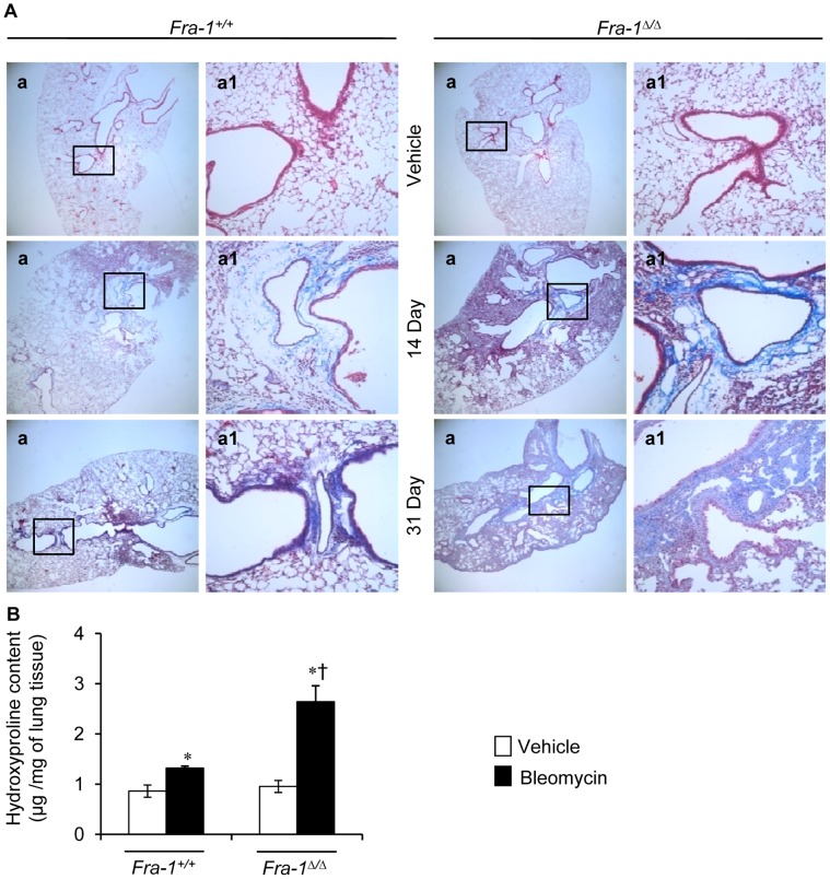
Figure 4. Fra-1 Δ/Δ mice develop exaggerated pulmonary fibrosis after injury. A:
Representative results of Masson’s trichrome staining of the lung from the saline-treated mice (n = 3) or bleomycin treated mice for 14 (n = 3) and 31 (n = 4) days. B: Right lung was collected for biochemical analysis of bleomycin-induced pulmonary fibrosis as measured by hydroxyproline content at 31-day post-PBS and -bleomycin treatment (n = 5). ∗p<0.05, PBS vs bleomycin; †p<0,05, Fra-1 Δ/Δ vs Fra-1+/+ mice. Images in a are shown at x4, whereas a1 represent boxed areas of a, shown at x20.

