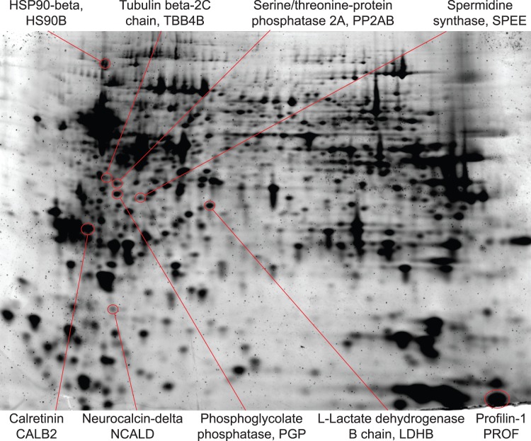
Figure 8. Proteins isolated from CTAS-dissected tissue samples preserve high integrity and may be used for downstream applications including 2D gel electrophoresis.
Representative 2D gel electrophoresis analysis of fresh frozen CA2–CA3 and DG areas of hippocampus (stained by Sypro Ruby) is shown. Protein samples from CA2–CA3 and DG areas were labeled with Cy3 and Cy5 correspondingly, imaged on the Typhoon, and analyzed using DIA module of Decyder. Gel was fixed and stained with Sypro Ruby; protein spots were picked and identified using MS. The following differences in protein abundances between identified proteins isolated by CTAS from CA2–CA3 and DG areas were observed (CA2–CA3/DG abundances ratios are shown in parenthesis): HS90B (−1.52), TBB4B (−2.34), PP2AB (−1.55), PGP (used as control, no difference), SPEE (−1.59), LDHB (used as control, no difference), CALB2 (1.76), NCALD (1.77), PROF1 (1.65).

