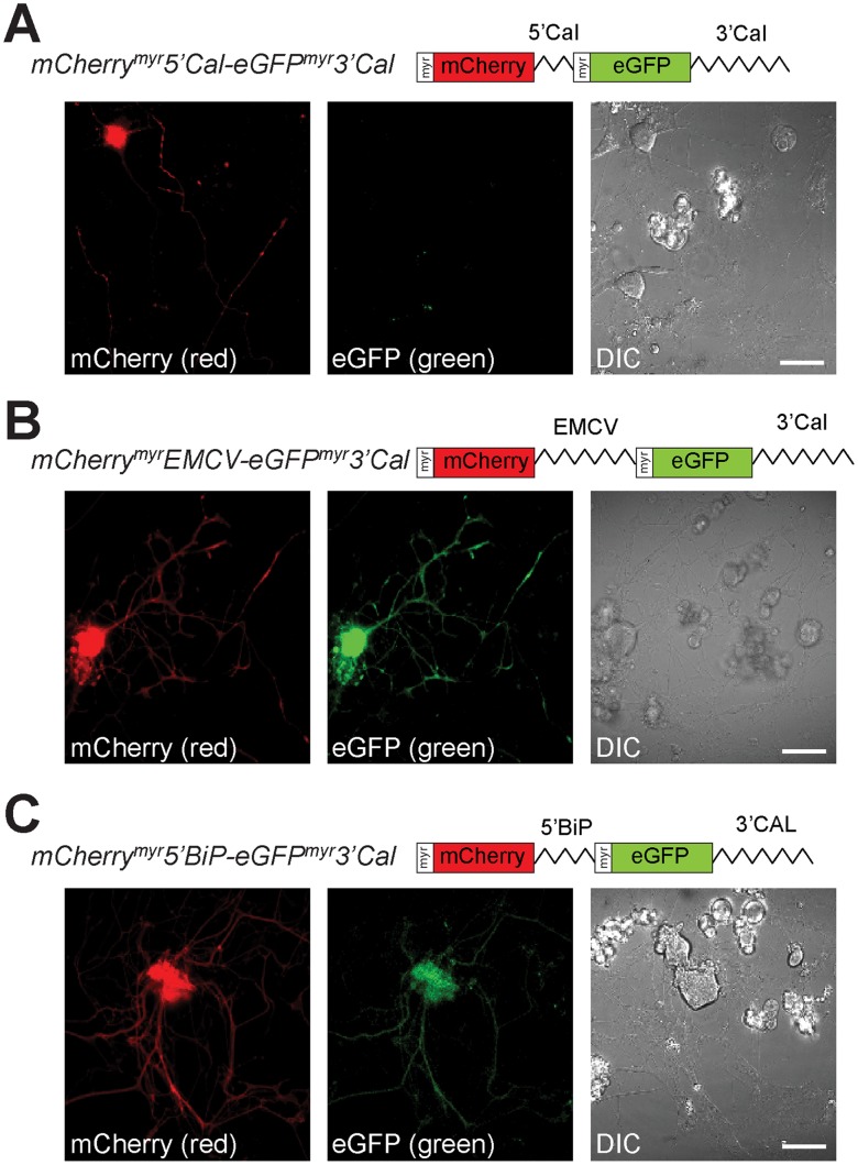
Figure 2. Bicistronic reporters support IRES-dependent translation in sensory neurons.
Representative static images of DRG neurons transfected with bicistronic pmCherrymyr5′Cal-eGFPmyr3′Cal (A), pmCherrymyrEMCV-eGFPmyr3′Cal (B), and pmCherrymyr5′BiP-eGFPmyr3′Cal (C) reporters are shown at 48 h post-transfection. Both mCherry (red, left panel) and eGFP (green, right panel) fluorescence is seen in the cell bodies and axons of mCherrymyrEMCV-eGFPmyr3′Cal and mCherrymyr5′BiP-eGFPmyr3′Cal expressing neurons (B,C), only the mCherry signals are seen for the mCherrymyr5′Cal-eGFPmyr-3′Cal expressing neurons (A). These data suggest that the 5′UTR of grp78/BiP mRNA but not calreticulin’s 5′UTR can function as an IRES in sensory neurons [scale bars = 50 µm].

