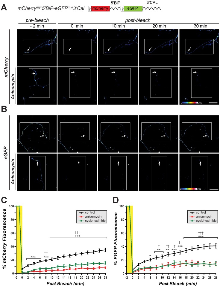
Figure 5. 5′UTR of grp78/BiP mRNA can function as an IRES in axons. A-B.
, Representative time-lapse sequences from FRAP analyses of DRG neurons transfected with pmCherrymyr5′BiP-eGFPmyr3′Cal as in Fig. 4 are shown. Sequences for cap-dependent translation of mCherry are shown in A and for IRES-dependent translation of eGFP are shown in B. The upper rows for A and B show cultures standard medium and lower rows show cultures pretreated with 150 µM anisomycin [scale bar = 50 µm]. C-D, Quantifications of axonal mCherry and eGFP signals from multiple FRAP sequences from DRG neurons transfected with mCherrymyr5′BiP-eGFPmyr-3′Cal are shown. Signals in each individual series are normalized to pre-bleach levels and are expressed as average percent prebleach signals ± SEM (n ≥6 over at least 4 independent transfections; *p≤0.05, **p≤0.01, and ***p≤0.001 for control vs. anisomycin time points and †p≤0.05, ††p≤0.01, and †††p≤0.001 for control vs. cycloheximide time points by two-way ANOVA compared to t = 0 min post-bleach). Both mCherry and eGFP fluorescence in distal axons shows recovery after photobleaching that is attenuated by protein synthesis inhibitors. These data indicate that the 5′UTR of rat grp78/BiP mRNA can drive IRES-dependent translation in axons.

