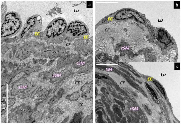
Figure 4. Morphological changes in lymphatic vessel endothelial cells.
Scanning electron microscopy (SEM) findings. (a) Lymphatic vessel endothelial cells protruding into the lymphatic lumen in the normal type. Bar = 10 µm. (b) Lymphangiectasia at its early stages. The lymphatic vessel endothelial cells are slightly flattened. Bar = 5 µm. (c) Medium-stage lymphangiectasia. The lymphatic vessel endothelial cells are markedly flattened. Bar = 10 µm. Lu; Lumen, EC; endothelium cells, sSM; synthetic smooth muscle cells, cSM; contractile smooth muscle cells, CF; collagen fibers.

