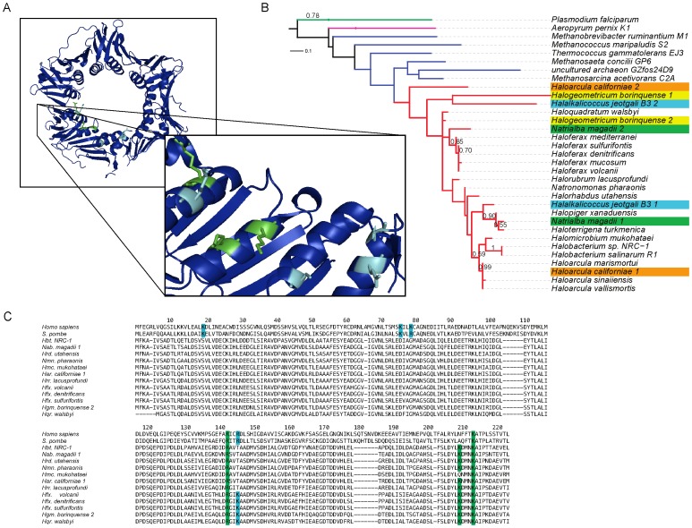
Figure 7. Proliferating cell nuclear antigen (PCNA).
(A) Crystal structure of Haloferax volcanii PCNA [61] with eukaryotic (light blue) and potential haloarchaeal DNA binding residues (green) shown. (B) Maximum likelihood tree of eukaryotic and archaeal PCNAs with bootstrap support values above 0.50 shown for 500 bootstrap iterations. Branch colors: green – eukarya, purple – crenarchaeota, dark blue – euryarchaeota, red - haloarchaea. Duplicate haloarchaeal PCNAs are distinguished with colored leaves. (C) An alignment of eukaryotic and haloarchaeal PCNA homologs. Residues known to be involved in DNA binding in eukaryotes are shown in light blue, with suspected functionally homologous positions in haloarchaea shown in green.

