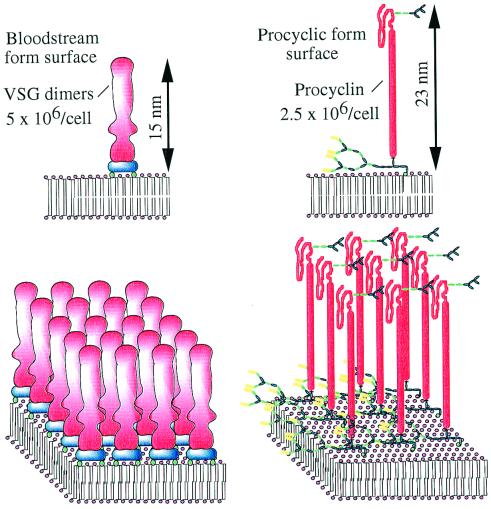
Figure 1.
The major surface molecules of T. brucei bloodstream and procyclic forms. The cartoons represent 20 nm × 20 nm portions of plasma membrane. The blue components of the VSGs represent the two GPI anchors that attach the VSG dimers to the membrane. The mature GPI anchors of the procyclins have very complex side chains and some procyclins contain small N-linked oligosaccharides beyond the polyanionic rod domain, as shown here.

