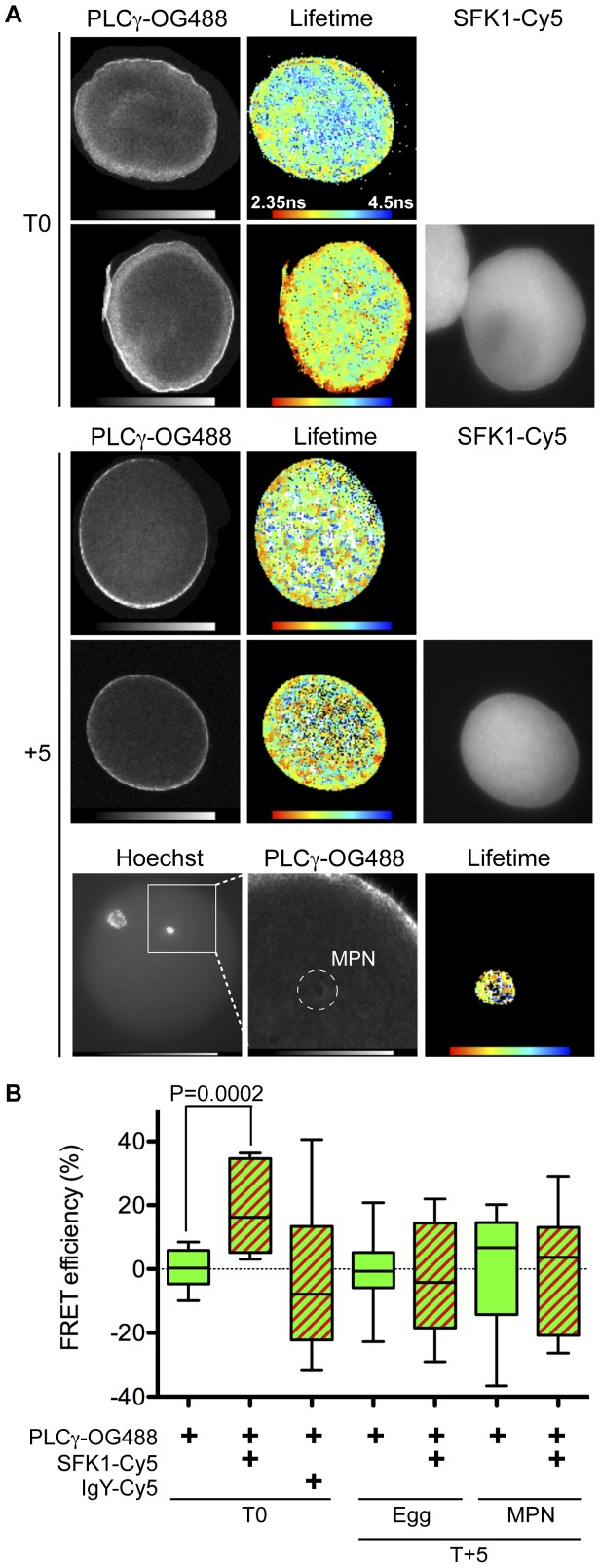
Figure 2. PLCγ and SFK1 interact during the early stages of male pronuclear envelope formation in vivo.
(A) Unfertilised L. pictus eggs (T0) or five minutes post-fertilisation (T+5) were fixed and labelled with anti-PLCγ-OG488 alone or together with anti-SFK1-Cy5. Samples were subjected to two-photon time domain FLIM and the lifetime of the donor chromophore (OG488) determined in the absence and presence of acceptor (Cy5). Donor (PLCγ-OG488) two-photon images, donor lifetime ‘heat maps’ and Hg-lamp epifluoresence acceptor (SFK-Cy5) images are shown. At T+5 the donor lifetimes in the whole egg (middle panel) and in the vicinity of the MPN only (bottom panel) were determined. In the donor two-photon image the MPN region of interest is indicated (dashed circle). The donor lifetime heat map and corresponding two-photon image are at the same scale. The images shown are from a donor alone labelled egg. The same process was repeated with eggs labelled with donor and acceptor. Data are representative of at least three independent experiments performed. (B) The FRET efficiency for each condition was calculated. Solid green boxes are the donor alone condition, green/red stripes for donor and acceptor conditions. Data are from at least three independent experiments, with a total of 6–18 eggs analysed per condition. Boxes display the median, upper and lower quartiles and whiskers the maximum and minimum values.

