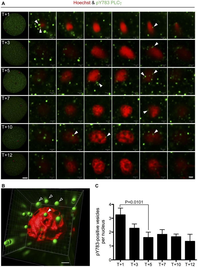
Figure 3. PLCγ is transiently phosphorylated on Y783 at the NE in vivo.
(A) Fertilised (T1–12) L. pictus eggs were fixed and stained with anti-pY783 on PLCγ, (green) and Hoechst (red), and imaged by confocal microscopy. Arrows (white) indicate pY783 positive vesicles recruited to the forming MPN. Data are representative of those obtained in three independent experiments. Scale bar is 20 µm (whole egg) or 1 µm (20× zoom). (B) Z-series from (A) were manipulated in Imaris (see Methods) to form a 3D reconstruction. Membrane vesicles in contact with the nucleus surface were scored (solid arrow). Those in close proximity were not (open arrow). (C) Quantification of the data in A, B. Vesicles were scored in three independent experiments. Data are expressed as mean+s.e.m.

