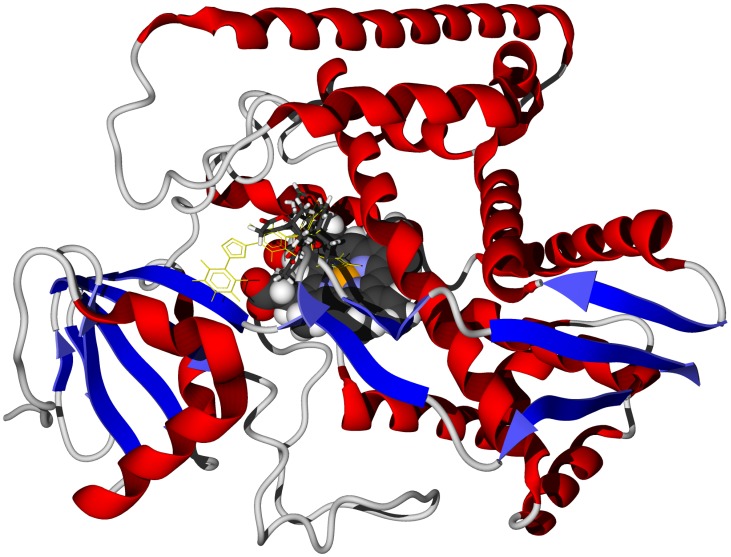
Figure 1. The crystal structure of T. brucei sterol 14α-demethylase, TbCYP51 (PDB 3gw9) [34].
The docked ligand is carapolide A (stick figure). The co-crystallized ligand is shown as a green wire figure and the heme cofactor is shown as a space-filling structure.

