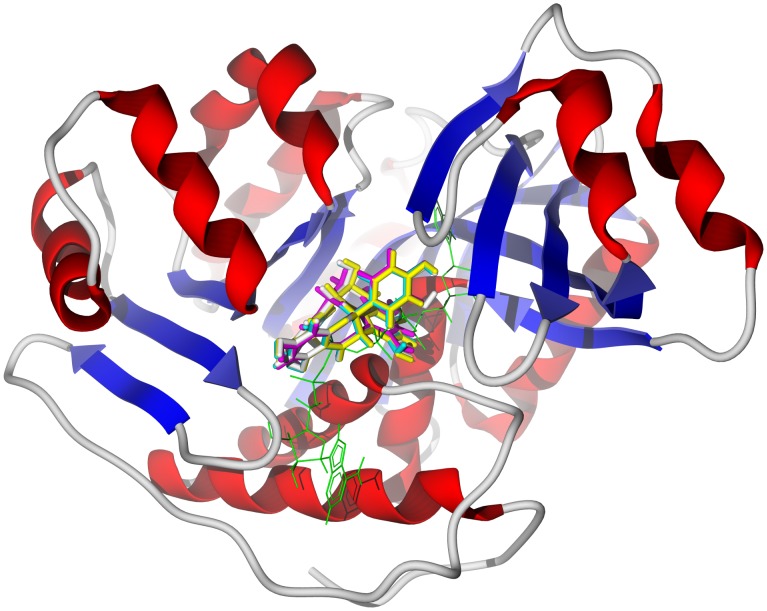
Figure 3. The crystal structure of T. brucei adenosine kinase, TbAK (PDB 3otx) [42].
The docked poses are the biflavonoids GB1 (turquoise), GB1a (magenta), GB2 (yellow), and garciniflavanone (white). The co-crystallized ligand, bis(adenosine)-5′-pentaphosphate, is shown as a green wire figure.

