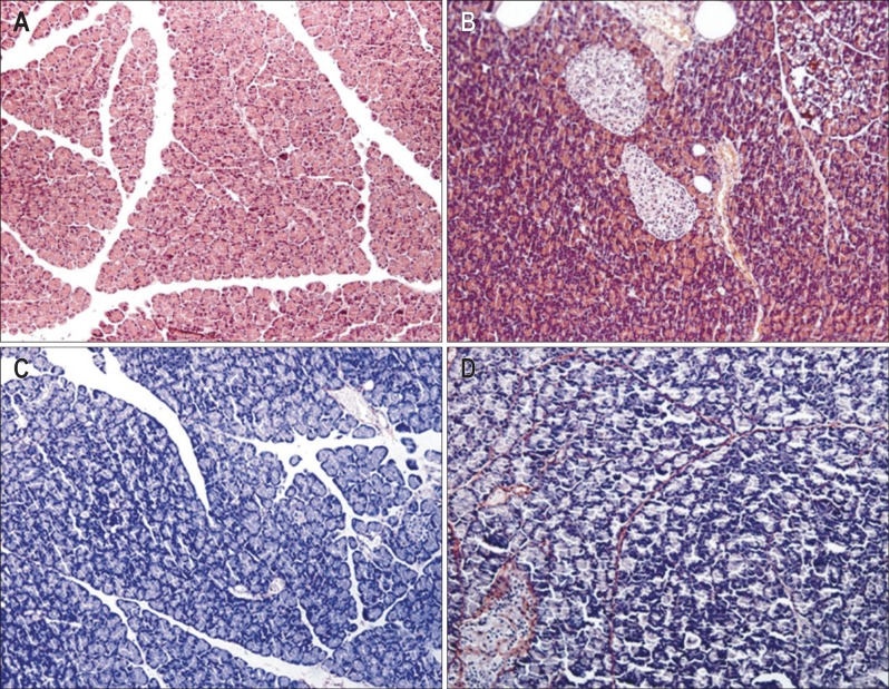
Fig. 1.
(A, B) Hematoxylin-eosin (H&E stain, ×200) and (C, D) Sirius red staining (×200) in pancreatic samples. The collagen fibrils appear red, and the non-fibrotic areas appear blue after Sirius red staining. The stained sections display none of the obvious histopathological changes observed in the control group (A, C), but fat deposition in acinar cells (cytoplasmic vacuolization), lymphocyte infiltration and collagen deposition were observed in the high-fat diet group (B, D), indicating the presence of fibrogenesis in the pancreas after prolonged ingestion of a high-fat diet. Control, n=10; High-fat, n=12.

