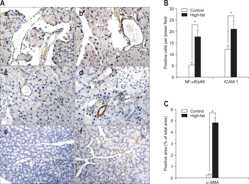
Fig. 2.
Immunohistochemical staining for nuclear factor kappa B p65 (NF-κB/p65), intercellular adhesion molecule 1 (ICAM-1), and α smooth muscle actin (α-SMA) (×400) and the corresponding statistical analysis. (A) Representative immunohistochemistry for NF-κB/p65 (a, b), ICAM-1 (c, d) and α-SMA (e, f) (control, a, c, e; high-fat, b, d, f). For NF-κB/p65 staining, cells with stained cytoplasm and a stained nucleus (brown) were considered positive. (B, C) The corresponding statistical analysis. The average number of NF-κB/p65- or ICAM-1-positive stained (brown) cells per high power field is reported. Areas staining positive for α-SMA are expressed as percentages of the total area. Data are presented as means±SD. Control, n=10; High-fat, n=12.
*p<0.001, control versus high-fat diet group.

