Fig. 1.
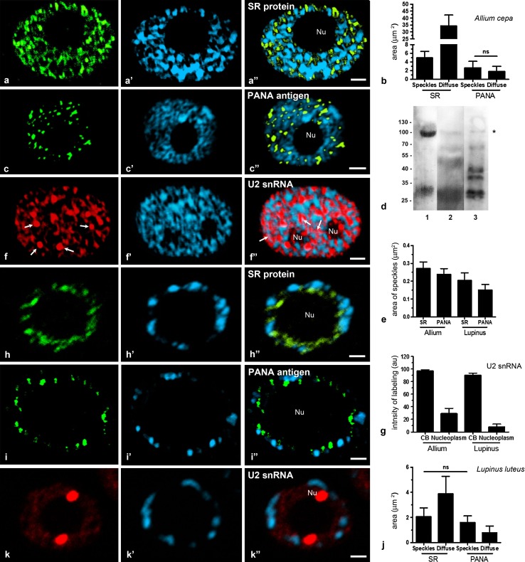
Localisation of SR proteins, PANA antigen and U2 snRNA in reticular (Allium cepa a, c, f) and chromocentric nuclei (Lupinus luteus h, i, k). a Localisation of SR proteins in the meristematic cells of roots. b The total area of nuclear structures similar to speckles and diffuse region stained with antibodies to SR proteins and to the PANA antigen in the Allium cepa nuclei. Ends of lines (ns) indicate regions of nucleus with nonsignificant difference. c Localisation of PANA antigen. There are structures resembling speckles in the nucleus of A. cepa. d Western blot of PANA antibody with total protein extracts from HeLa cells (1), nuclei of roots Allium cepa (2), and nuclei of roots Lupinus luteus (3). e The size of nuclear structures stained with antibodies to SR proteins and to the PANA antigen in the A. cepa and L. luteus nuclei. f Localisation U2 snRNA. The bright spots are Cajal bodies (arrows). Diffuse staining of the nucleoplasm is also observed. f″ Co-localisation of signals from U2 snRNA and DNA staining with DAPI were observed (arrows pink in colour). g The intensity of labelling in CB and nucleoplasm stained with U2 probe. Nuclear localisation of SR proteins (h) and PANA antigen (i) in a chromocentric nucleus. j The intensity of labelling of speckles and diffuse nuclear fraction stained with antibodies to SR proteins and to the PANA antigen of L. luteus. Ends of lines (ns) indicate regions of nucleus with nonsignificant difference. k There are two Cajal bodies and a diffuse pool stained U2 probe in the nucleus of L. luteus. a′, c′, f′, h′, i′, k′ DAPI staining. a″,b″,f″,h″,i″,k″ Overlay DAPI staining and corresponding antigens. Nu nucleolus. Bar 5 μm

