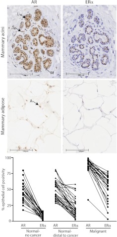
Fig. 1.
AR and ERα in human breast tissues. Panel A, Representative images of immunostaining for AR (left panel) and ERα (right panel) in acini of human breast tissue. Luminal epithelial (L), myoepithelial (M), and stromal (S) cells are indicated. Panel B, Representative images of immunostaining for AR (left panel) and ERα (right panel) in breast adipose tissue. An AR-positive adipose (A) nucleus is indicated by the arrow. Panel C, Percentage of AR- and ERα-positive epithelial cells in prospectively collected breast tissues representing normal breast tissues from women without breast cancer (n = 23), normal breast tissues distal to a malignancy (n = 38), and malignant breast tumor tissues (n = 27).

