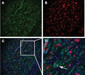
Fig. 5.
Colocalization of AR and Ki67 in an ERα-negative breast carcinoma. A–C, Representative images of fluorescent immunostaining for AR (green) (A), Ki67 (red) (B), and both antigens merged with DAPI (blue) (C) in an ERα-negative breast carcinoma; D, higher-power image of boxed area in C showing an AR-positive, Ki67-positive tumor cell (white arrow).

