
Fig. 7.
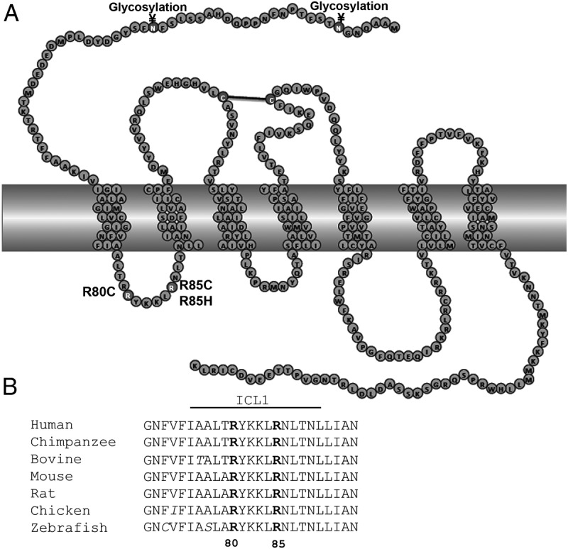
PROKR2 ICL1 amino acid sequence alignment among species. A, Schematic representation of mutations in the ICL1 of PROKR2. Residues in dark gray represent the ICL1 mutations. Residues involved in disulfide bond formation (black line) and in glycosylation are represented in gray. B, Amino acid sequence alignment among species, showing that arginines in position 80 and 85 are highly conserved among different species. Sequences were taken from the GPCR database (www.gpcr.org), and the alignment was produced in ClustalW.