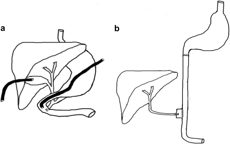
Fig. 1.
A, Schematic illustration of the anatomy and canulation of the canine model. A gastrostomy tube was placed into the duodenum close to the ampulla of Vater. The common bile duct was ligated and the gallbladder canulated to allow drainage of bile. B, Schematic illustration of the functional anatomy of the bile in ileum group. Transections 1 cm proximal and distal to the drainage point of the common bile duct were performed. The proximal and distal ends of the transected duodenum were anastomosed end to end and continuity restored. The segment of the duodenum containing the common bile duct was anastomosed side to side to the distal jejunum, 10 cm proximally to the terminal ileum.

