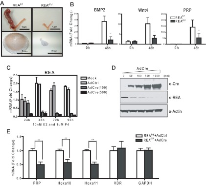
Fig. 3.
REAd/d mice show impaired uterine decidual response in vivo and in vitro. A, Representative gross anatomy of uteri from REAf/f and REAd/d mice after decidual stimulation. The left horn was stimulated, and the right horn was not stimulated (see Materials and Methods). Note the dramatic increase in size of the left horn of REAf/f uteri after stimulation. Lower panels show REA in the stimulated REAf/f left horn by immunohistochemistry (IHC). Note high expression of REA in the decidual zone. B, Expression of molecular markers for decidualization measured by qRT-PCR at 0 h and 48 h after stimulation. C, In vitro decidual response of uterine stromal cells. Mouse stromal cells from d-4 pregnant uteri were infected by mock or control adenovirus or different multiplicity of infection (MOI) of AdCre, and then exposed to 10 nm E2 and 1 μm P4 for times up to 96 h. REA mRNA (C) and protein levels (D) were then analyzed by qRT-PCR (24–96 h) and immunoblotting (96 h), respectively. E, Molecular markers specific to stromal cells were monitored at 96 h by qRT-PCR. Data are mean ± sd. *, P < 0.05; **, P < 0.01. Wnt4, Wingless-related MMTV integration site 4; PRP, prolactin-related protein; VDR, vitamin D receptor; GAPDH, glyceraldehyde-3-phosphate dehydrogenase.

