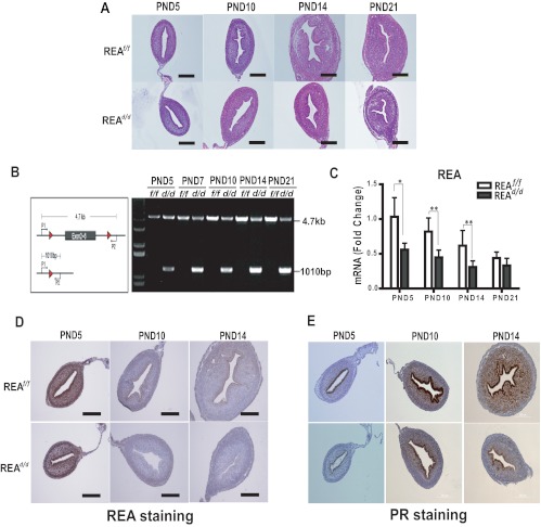
Fig. 4.
REAd/d mice show altered uterine growth and maturation. A, Uterine morphology was examined in hematoxylin- and eosin-stained sections from uteri of REAf/f and REAd/d mice at PND5, PND10, PND14, and PND21. B, REA gene is excised in the uteri of REAd/d mice from as early as age 5 d (PND5). REA deletion was confirmed by genotyping. C, REA mRNA level was monitored by qRT-PCR in uteri from PND5, PND10, PND14, and PND21. Values are mean ± sd (n = 10 per group), and mRNA levels are illustrated as relative expression normalized to 36B4 by wild-type PND5. *, P < 0.05; **, P < 0.01. Immunohistochemical detection of REA (D) and PR from PND5, PND10, and PND14 uteri of REAf/f and REAd/d mice (E). Magnification is the same for D and E. Scale bar, 200 μm.

