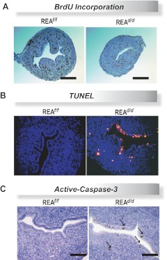
Fig. 6.
REAd/d mice have decreased neonatal uterine cell proliferation and increased apoptosis. A, Representative histologic sections of BrdU immunohistochemistry in uteri of REAf/f and REAd/d d-14 mice. Note decreased BrdU incorporation in the REAd/d uterus. Scale bar, 200 μm. B, Representative fluorescence images of TUNEL staining in uterine sections from REAf/f and REAd/d d-14 mice. C, Immunohistochemical detection of active-caspase-3 in uteri of REAf/f and REAd/d d-14 mice. Scale bar, 100 μm.

