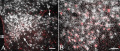
Fig. 7.
Colocalization of c-Fos and VGLUT2 in the PVN of animals refed for 2 h after fasting. Silver grains denoting the VGLUT2 mRNA are located over all c-Fos-IR neurons (red) in both the PVNv (vp) and PVNl (lp) (A). Higher-magnification image illustrates the colocalization of c-Fos and VGLUT2 mRNA in the PVNv (B). III, Third ventricle; lp, lateral parvocellular subdivision; vp, ventral parvocellular subdivision. Scale bar on A, 100 μm; scale bar on B, 50 μm.

