Fig. 2.
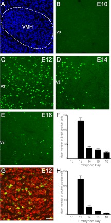
Embryonic birthdate of neurons in the VMH. A, Confocal image illustrating nuclear (bis-benzamide) staining used to recognize the morphological limits of the VMH. B–E, Confocal images showing the presence of BrdU (green)-positive cells in the VMH of P10 mice that were injected with BrdU at E10 (B), E12 (C), E14 (D), or E16 (E). F, Quantification of the numbers of BrdU-positive cells in the VMH of P10 mice that were injected with BrdU at E10, E12, E14, E16, or E18 (n = 4–6 per group; three sections per animal). G, Confocal image showing the presence of HuC/D immunoreactivity (a neuronal marker, red) in VMH BrdU (green)-positive cells of a P10 mouse that was injected with BrdU at E12. H, Quantification of the numbers of BrdU+HuC/D double-labeled cells in the VMH of P10 mice that were injected with BrdU at E10, E12, E14, E16, or E18 (n = 4–6 per group; three sections per animal). V3, Third ventricle. Values are the means ± sem. Scale bar, 50 μm (A–E) and 30 μm (G).

