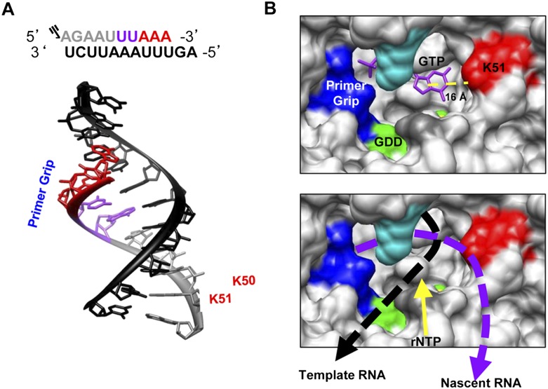
FIGURE 10.
A model for the paths of the nascent and template RNAs as they exit the HCV RdRp. (A) Schematic and structure of 5P and 12T relative to the primer grip and residues K50 and K51. The structure shown is an A-form RNA helix, trimmed to match the size of 12T and 5P (PDB: 1RNA). (Gray) 5P; (black) 12T; (purple and red residues) the walking of the polymerase. (B, upper panel) Regions and structural features within NS5B to contact the nascent RNA. The location of the initiation GTP was used to denote the position of the first nucleotide that could form the nascent RNA. (Yellow sphere) The Mg2+ ion that coordinates the GTP to the GDD active site (green). (Red) Residue K51 that forms the beginning of the nascent RNA channel. The distance between C5 of the guanine to the terminal ɛ-amino atom of K51 is shown below the yellow dashed line. (B, lower panel) The proposed paths of the nascent and template RNA. The models used the coordinates in the polymerase–GTP complex of Harrus et al. (2010) (PDB: 2XI3). The figures were drawn with the Chimera program (UCSF, San Francisco) (Pettersen et al. 2004), and labels and features within the polymerase were added using Microsoft PowerPoint.

