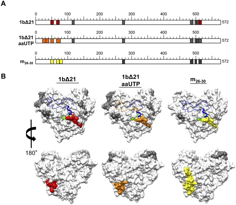
FIGURE 3.
Mapping of the residues in the HCV RdRp that contact the nascent RNA. (A) Schematic representation of peptides identified in the RCAP assay variations. Colored regions represent peptides identified during RNA synthesis: (red) regions identified by the RCAP assay of 1bΔ21; (orange) regions identified during RNA synthesis using a nucleotide analog, aminoallyl-UTP (aaUTP); (yellow) regions identified within m26-30, an RdRp mutant defective in de novo initiation. Details regarding the peptide assignments can be found in Supplemental Figure 2. The reactions were performed as described in Materials and Methods. Peak assignments to peptides within NS5B were based on theoretical digests that match calculated masses within 0.5 Da. (B) Molecular model of the peptides within NS5B found to contact RNA (PDB: 1QUV). (Gray) Peptides found to contact 5P or 5P/12T; ones present only with the ternary complex generated in the presence of UTP (red), UTP (orange), or ATP (yellow); (green) the active site of NS5B; (blue in the ribbon structure) the Δ1 loop.

