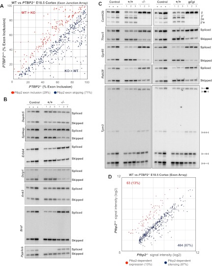
Figure 2.
Ptbp2-dependent alternative splicing in the brain. (A) Relative exon inclusion levels in wild-type (WT) and Ptbp2 knockout (KO) brains (ASPIRE3, P < 0.01, ΔIrank > 5). Red dots indicate alternative exons with higher inclusion in wild-type compared with knockout brains, and blue dots represent exons with increased inclusion in knockout compared with wild-type brains. (B) RT–PCR validation of seven ASPIRE candidates (additional candidates in are shown in the Supplemental Figures). In all cases, the control sample consists of an equal-parts mixture of wild-type and knockout cDNA amplified at different cycle numbers (see the Materials and Methods). (C) Comparison of alternative splicing in both Ptbp2-null strains (−/−, gene targeting; gt/gt, gene trap) (for additional examples, see Fig. 6). (D) Probeset signal intensities from MoEx 1.0ST analysis of wild-type and Ptbp2-null brains.

