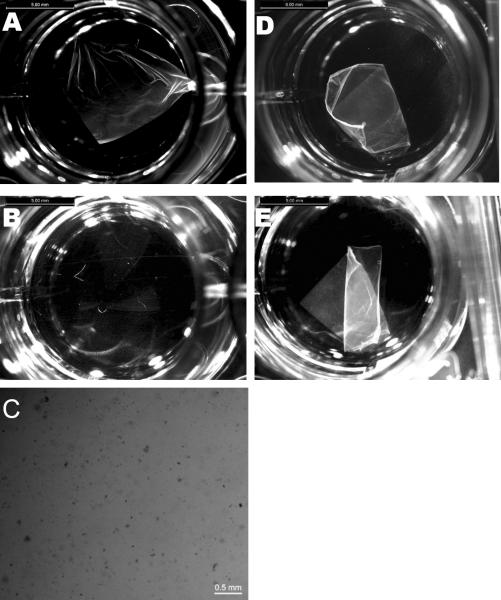
Figure 5.
Bright field microscopy of electrospun honeybee silk mats. Mat before adding α-chymotrypsin (A), and same mat after 24 h incubation in α-chymotrypsin (B). Commassie Blue stained fragments of mat after α-chymotrypsin digestion (C). Control mat in PBS at t=0 (D), and same mat after 24 h (E). Scale bars: 2 mm.

