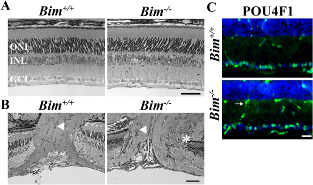
Figure 1. Bim deficiency affects retinal development.

(A) Retinal sections from Bim+/+ and Bim-/- mice confirms previous reports1,17 that Bim deficiency did not alter the gross organization of number of retinal neurons. (B) Bim deficiency caused dysmorphogenesis of the optic nerve. In Bim deficient mice the retinal-optic nerve head border is abnormal, with apparent retinal neuronal layers entering the optic nerve head (asterisk). Also the normal arrangement of glia cell bodies does not appear to be present in the area of the lamina cribrosa (arrowhead). (C) However, optic nerve head morphology changes did not affect the number of RGCs, as judged by POU4F1 (BRN3A, green) expression, which is specifically expressed in 80% of RGCs in the retina19. Note, the secondary antibody also detects retinal vasculature (arrow). DAPI, blue; ONL, outer nuclear layer; INL inner nuclear layer, GCL, ganglion cell layer; scale bar, A,B = 50 μm, C = 25 μm.
