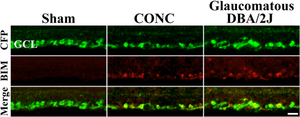
Figure 2. BIM is expressed in RGCs after axonal injury.

In young uninjured RGCs (Sham), BIM is not detected in RGCs. RGCs are marked genetically with CFP using B6.Thy1-CFP mice. In these mice 95% of RGCs express CFP and only a small percent of the other type of neurons in the ganglion cell layer, displaced amacrine cells, express the transgene20,21. At the beginning of RGC death after CONC (3 days after injury) BIM colocalizes with the vast majority of CFP+ cells. BIM continues to be expressed in the RGC layer at 5 days and 7 days indicating it is expressed throughout the time when RGC death peaks. In glaucomatous DBA/2J mice, BIM is also expressed in RGCs (RGCs are also marked with the Thy1-CFP transgene; backcrossed into DBA/2J mice >20 times). BIM expression was not detected in all sections, but was present in RGCs in 4 out of 6 retinas examined. This expression is consistent with the asynchrony of DBA/2J glaucoma, with only some 10 month old animals undergoing active RGC loss29. Also, in diseased retinas, BIM was not detected in every section likely reflecting the naturally-occurring sectorial pattern of RGC loss8. In addition, BIM expression was absent in 10 month old D2.Gpnmb mice (data not shown; D2.Gpnmb mice are a control strain of D2 mice that are wild-type for one of the genes that causes the iris disease and do not develop elevated IOP40,44). The lack of BIM expression in D2.Gpnmb RGCs indicates that change in BIM expression in DBA/2J RGCs is caused by the glaucomatous insult and not age or genetic background. These data show that BIM is expressed in axonally injured RGCs. Scale bar, 25 μm.
