
Figure 4. Evidence for Nrf2 antioxidant program in pancreatic cancer.
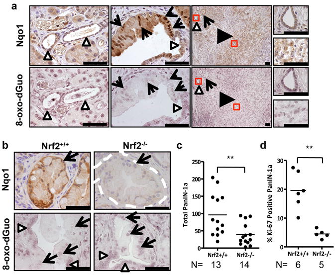
a, Immunohistochemical detection of Nqo1 (brown staining) and 8-oxo-dGuo (purple staining) in mouse PanIN and PDA (similar patterns observed for 11/11 of cases examined) in comparison to morphologically normal ducts. PanIN (arrows), PDA (black arrowheads), normal ducts (white arrowheads) here and for all figures. Scale bar = 56 μm. b, Immunohistochemical detection of Nqo1 and 8-oxo-dGuo in Nrf2-/- PanIN compared to Nrf2+/+ PanIN (similar patterns observed for 5/5 of each genotype examined, PanIN outlined by white dashes). Scale bar = 56 μm. c, Nrf2-/- and Nrf2+/+ PanIN-1a incidence. Whole pancreata were sectioned at 100-micron intervals and total numbers of PanIN-1a were counted. d, Proliferation of PanIN-1a cells in Nrf2-/- and Nrf2+/+ mice, as determined by Ki-67 immunostaining.