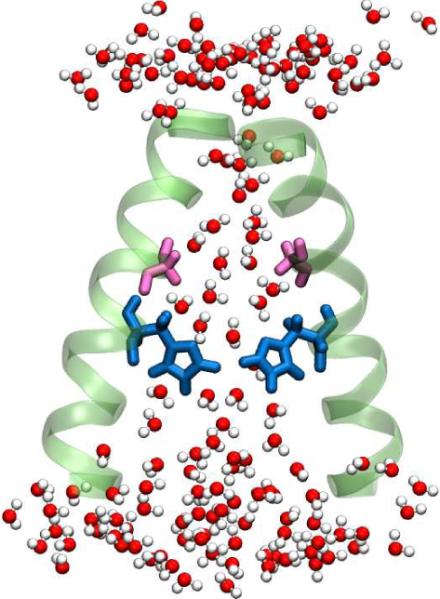
Figure 6.

A cartoon of the M2 channel, showing the water structure inside the channel. The number of water molecules accessible to the pore lining residues is significantly less than in bulk solution. The above picture was generated from MD simulation trajectories performed with the 3LBW crystal structure. Only two helices are shown for clarity. The Gly34 residues are shown in pink and the His37 are shown in blue.
