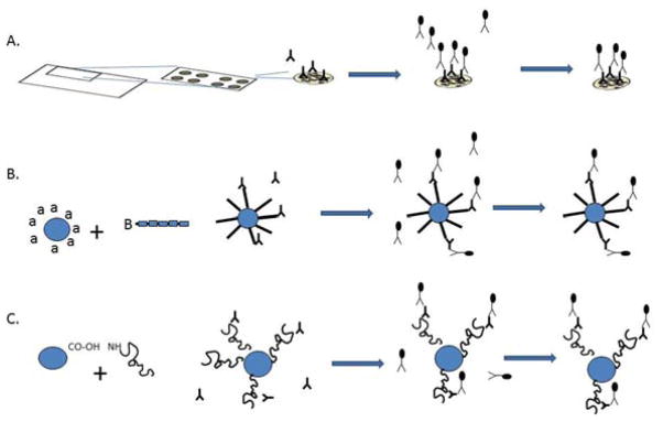
Figure 1.
Schematic representation of planar and bead-based microarrays. A. Planar antigen microarrays, B. Bead-based peptide antigen array, C. Bead-based protein antigen array. In all cases, antigen is bound to the experimental surface, probed with antibody-containing fluid sample, washed, incubated with secondary detection antibody, and visualized for quantitation of primary antibody reactivity.

