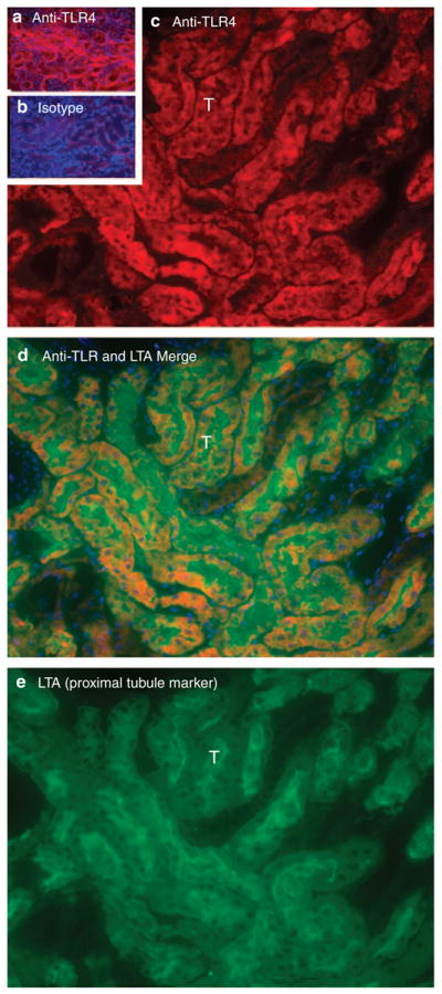
Figure 10. Proximal tubular Toll-like receptor 4 (TLR4) at 24 h reperfusion.

(a) Outer stripe of the outer medulla (OSOM) stained for TLR4 (red). (b) OSOM stained with isotype control primary antibody. Exposure times for a and b are identical. (c) High-power view of OSOM stained for TLR4. ‘T’ is one of many tubules in c–e. (d) Merged lotus tetragonolobus lectin (LTA) and TLR4 of same section. Blue is 4,6-diamidino-2-phenylindole (DAPI) staining of nuclei. DAPI staining reveals nuclei. LTA marks proximal tubule cells. (e) High-power view of same section stained for LTA.
