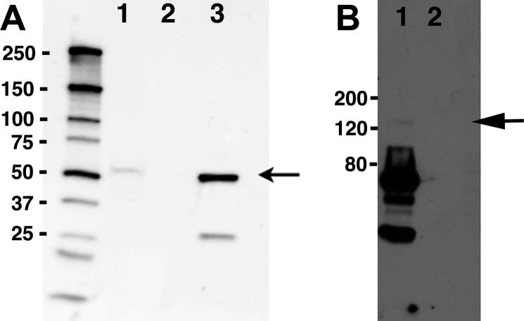
Fig. 2.
Reciprocal immunoprecipitation experiments confirm an interaction between cSlo and Rack1. Rack1 was immunoprecipitated with Rack1 (B3) antibody (Santa Cruz) from lysates of a stable cell line expressing FLAG-cSlo-yellow fluorescent protein (YFP) transfected with Rack-1-CFP. Slo was immunoprecipitated using FLAG M2 antibody (Sigma). The immunoprecipitates were separated on SDS-PAGE gels, and the reciprocal protein was detected by Western blotting. A: imunoprecipitates of FLAG contain Rack1-CFP [arrow 62 kDa (35 kDa RACK1 + 27 kDa CFP)] detected using anti-Rack1 antibody (lane 1). Immunoprecipitates using mouse serum served as a negative control (lane 2). The lysates from stable cell line expressed FLAG-cSlo-YFP (lane 3) and served as the positive controls. The molecular weight marker sizes are shown on the left. B: immunoprecipitates of Rack contain FLAG-cSlo-YFP (arrow) detected using anti-FLAG M2 antibody (Sigma) (lane 1). Immunoprecipitates using mouse serum served as a negative control (lane 2). Protein molecular weight markers are indicated on the left.

