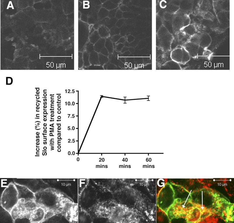
Fig. 6.
Increased Slo surface expression in response to PKC activation results in part from increased recycling of endocytosed Slo. To assess the recycling of internalized cSlo channels, the FLAG-cSlo-YFP stably expressing cells were incubated with anti-FLAG antibody for 1 h to allow labeled channels to undergo internalization. The surface-bound antibody was removed with stripping, and the cells were then reintroduced back in the culture medium ± PMA for varying period(s) to allow the recycling of the antibody-labeled internalized (endocytosed) channels. The cells were fixed with paraformaldehyde. The surface (recycled) cSlo channels were then labeled with secondary AB conjugated to Alexa 647 (Invitrogen). A–C: shown are confocal images of HEK cells expressing FLAG-cSlo-YFP that were subject to a recycling assay and demonstrate recycled Slo on the surface of the cell. Cells treated with PMA for 20 min show an increase in recycled Slo (C), compared with comparable control cells at time 0 (A) and at 20 min (B). D: presence of recycled Alexa 647 labeled cSlo channels were quantified by FACS. Shown is the percentile increase in surface labeled cSlo in PMA-treated cells compared with controls. There was an increase in recycled Slo molecules delivered to the surface of the cell upon treatment with 15 nM PMA compared with control at 20 min that sustained itself for over 1 h (mean fluorescence ( ± SE). E–G: shown are confocal images of HEK cells expressing FLAG-cSlo-YFP that were treated with transferrin (50 μg/ml) for 60 min. Cells were fixed and analyzed by confocal imaging. Slo-YFP (E) partially colocalizes with transferrin (F) with the merged pseudo-colored images shown in G (Green-Slo, Red-transferrin, and colocalized proteins in yellow). Arrows indicate internalized transferrin receptor. The images were obtained in the midplane of the cell.

