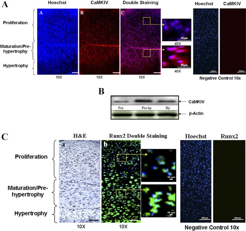
Fig. 2.
The distribution of CaMKIV and runt-related transcription factor-2 (RunX2) in the growth plate. To determine the distribution of CaMKIV and RunX2 in the growth plate, immunofluorescent histochemistry was performed with 5-μm-thick sections of the proximal tibia growth plate (from 1 day postpartum mice) using anti-CaMKIV and RunX2 antibodies. A: strong staining for the CaMKIV is found in the maturation/prehypertrophic zone compared with the proliferation zone. The boxed region in Ac, left, is represented at a higher magnification in Ac, right. Scale bar, 100 μm. Blue, nucleus; red, HDAC4; purple, overlap of blue and red. Normal goat IgG was used in place of CaMKIV primary antibody as negative control. No positive staining was detected for CaMKIV in the negative controls. B: to further validate the distribution of CaMKIV, protein extracts were obtained from the different zones of the growth plate. A similar expression pattern of CaMKIV is detected by Western blotting with anti-CaMKIV antibody. Hy, hypertrophy. C: RunX2 is strongly expressed in the prehypertrophic and hypertrophic zones with a weaker staining found in the proliferation zone. The boxed region in Cb, left, is represented at a higher magnification in Cb, right. Normal rabbit IgG was used in place of RunX2 primary antibody as negative control. No positive staining was detected for RunX2 in the negative controls. Scale bar, 100 μm. Blue, nucleus; green, RunX2. H&E, hemotoxylin and eosin.

