Fig. 6.
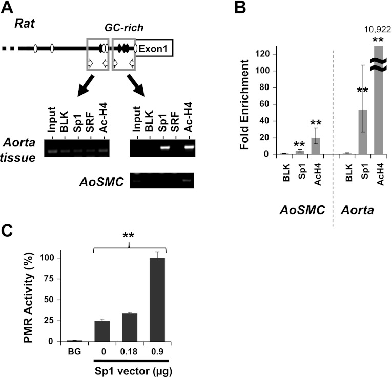
Binding of Sp1 to CPI-17 promoter. Chromatin immunoprecipitation (ChIP) assay was performed using rat aorta tissues and AoSMC with antibodies listed, followed by Conventional PCR (A) using two sets of primers for distal (left) and proximal (right) region of the CPI-17 promoter, and qPCR analysis for the proximal region (B). Preimmune IgG was used as blank and the ChIP signal was normalized against blank and shown as fold enrichment. **P < 0.05, compared with blank (n = 6). C: promoter assay (PMR) was performed using S2 cells transfected with the reporter vector for the mouse CPI-17 173-bp segment in the presence of the Sp1 vector at the indicated amount. Background (BG) was determined using the empty vector. **P < 0.05, by one-way ANOVA analysis (n = 3).

