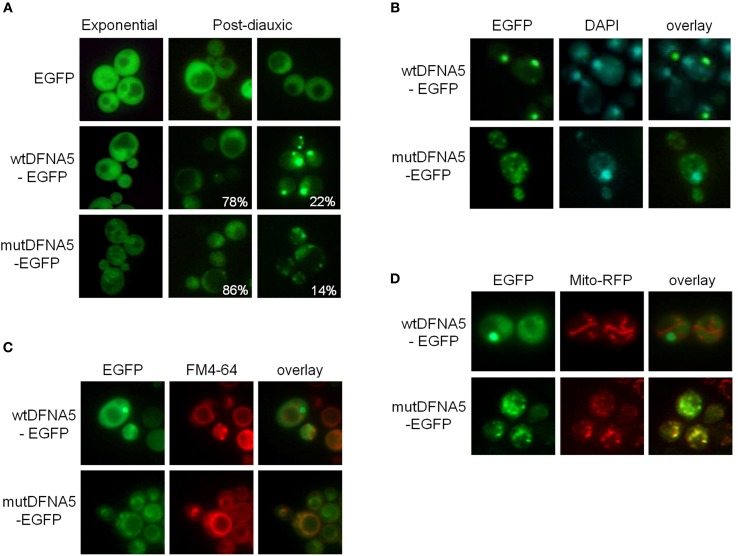
Figure 2.
Wild-type and mutant DFNA5 form inclusions in yeast. (A) Fluorescence microscopic visualization and intracellular localization of wtDFNA5-EGFP and mutDFNA5-EGFP fusion proteins in the BY4741 wild-type yeast strain sampled at the mid-exponential growth phase or in the post-diauxic growth phase as indicated. Percentages refer to the number of cells displaying a dispersed cytoplasmic localization or with inclusions. Cells expressing only EGFP served as control. (B,C) Pictures of post-diauxic wild-type cells with inclusions formed by wtDFNA5-EGFP or mutDFNA5-EGFP and stained with DAPI (panel B) to visualize the nucleus or with FM4-64 (panel C) to visualize the vacuolar membrane. (D) Pictures of post-diauxic wild-type cells expressing wtDFNA5-EGFP or mutDFNA5-EGFP and a mitochondrial red fluorescent marker protein, Mito-RFP.

