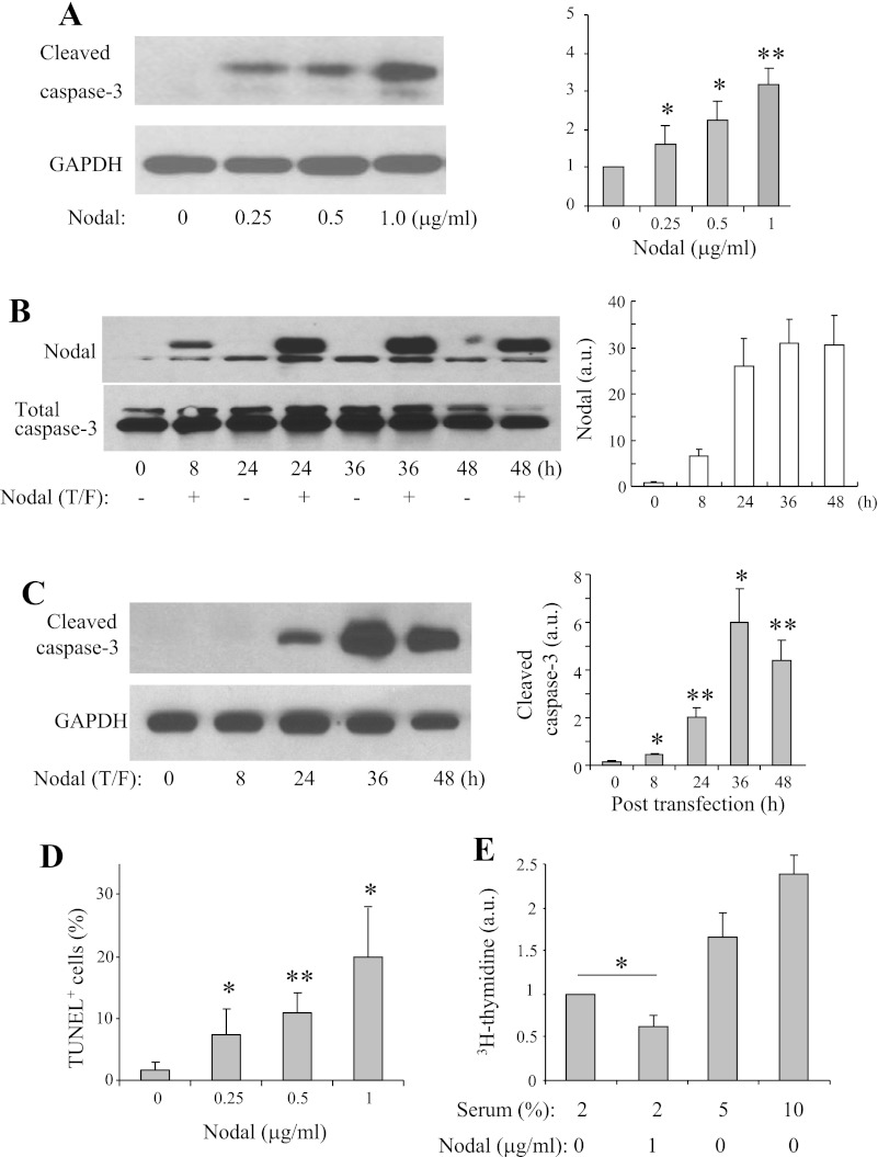
Fig. 4.
Nodal induces apoptosis and suppresses proliferation in INS-1 cells. A: Western blot analysis was performed in INS-1 cells treated with medium alone or with Nodal as indicated for 16 h. B: Western blotting detection of Nodal expression in INS-1 cells transfected with Nodal cDNA for indicated time. C: Western blot analysis was performed in INS-1 cells transfected with Nodal cDNA for indicated time. D: INS-1 cells were incubated with medium alone or with Nodal as indicated for 16 h, and apoptosis was quantified by TUNEL labeling. E: [3H]thymidine incorporation assay performed in INS-1 cells treated with or without Nodal (1 μg/ml) in the presence of 2% fetal bovine serum for 24 h (5 and 10% serum were used as positive controls). Data represent means ± SE; n = 3–5. *P < 0.05, **P < 0.01.

