Fig. 5.
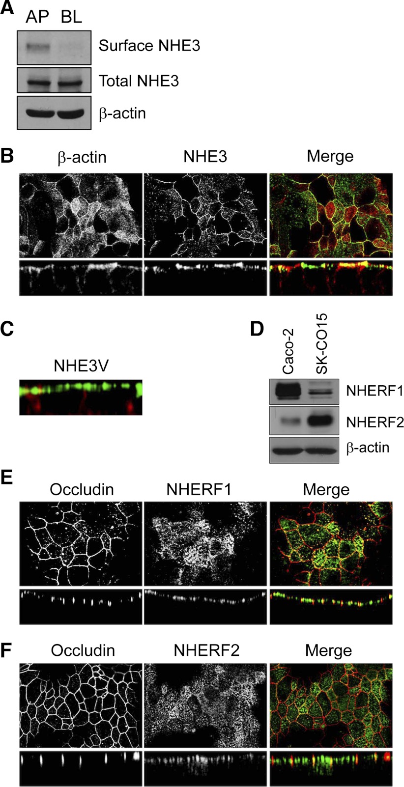
NHE3 is localized on the apical membrane in SK-CO15 cells. A: surface biotinylation was performed to determine the polarity of endogenous NHE3 expression in SK-CO15 cells as described in Surface biotinylation. Western blot analysis shows protein expression in the apical (AP) and basolateral (BL) fractions. β-actin was used as a loading control (n = 3). B: cells were fixed with ice-cold ethanol, permeabilized, and incubated with affinity-purified anti-NHE3 antibody EM450 followed by incubation with FITC-conjugated goat anti-rabbit secondary antibody. Rhodamine-conjugated phalloidin was used to label F-actin. Top and bottom: focal and cross-sectional views, respectively. C: cells transfected with pcDNA3.1/NHE3V were labeled with anti-vesicular stomatitis virus glycoprotein antibody P5D4 (green) and phalloidin (red). A cross-sectional view is shown. D: presence of NHE regulatory factor 1 (NHERF1) and NHERF2 in SK-CO15 cells was determined by Western blot analysis using the anti-NHERF1 antibody Ab5199 and anti-NHERF2 antibody Ab2570. β-Actin was used as a loading control. NHERF1 (E, green), NHERF2 (F, green), and occludin (E–F, red) were labeled with anti-NHERF1, anti-NHERF2, and anti-occludin antibodies, respectively. Top and bottom: show focal and cross-sectional views, respectively.

