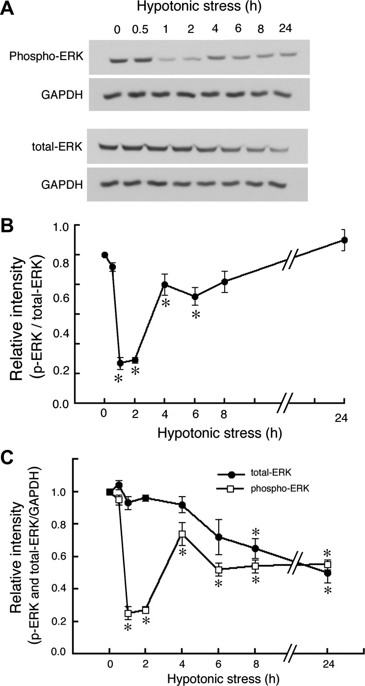
Fig. 2.
Effects of hypotonic stress on dephosphorylation of ERK. A6 monolayers were exposed to a hypotonic culture medium for the indicated times, and then ERK and phospho-ERK were detected by immunoblotting with anti-ERK and anti-phospho-ERK antibodies. A: typical immunoblots containing 45 μg whole-cell lysates. These blots were initially blotted with either anti-phospho-ERK or anti-ERK antibodies and then stripped and reprobed with GAPDH antibody. B: time-dependent changes in relative amounts of phosphorylated and total ERK proteins in response to hypotonic stress are shown. Equal loading of each well was confirmed by measuring GAPDH as an internal control (C). Values are means ± SE; n = 3. *P < 0.05.

