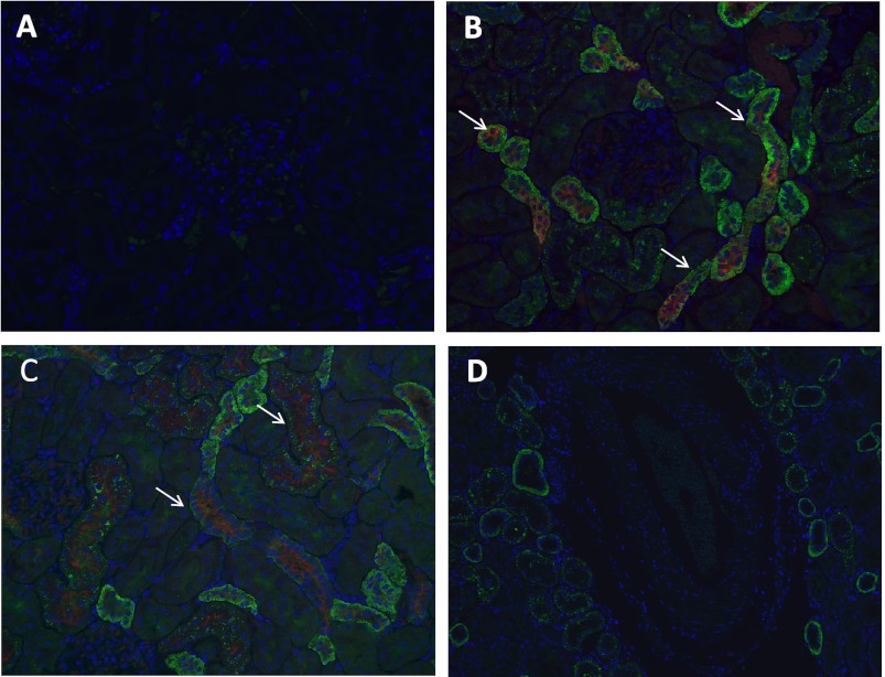
Fig. 2.
Cortical localization of the α7-nAChR. A: control slide incubated with primary antibody but without secondary antibody (×20). B: representative photomicrograph demonstrating expression of α7-nAChR in the proximal tubules as identified by Lotus tetragonolobus lectin (×20). C: representative photomicrograph of a slide showing expression of α7-nAChR in the distal tubules as identified by peanut agglutinin (×20). D: representative photomicrograph demonstrating lack of expression of the α7-nAChR in the intrarenal vasculature (×20).

