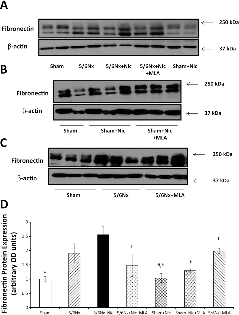
Fig. 5.
Cortical fibronectin expression. A, B, and C: representative Western blot for fibronectin and β-actin, which was used to control for loading. D: densitometry data analysis for cortical fibronectin expression (n = 6–7 per group; *P < 0.05 vs. 5/6 Nx and 5/6Nx + Nic; #P < 0.05 vs. 5/6Nx; †P < 0.05 vs. 5/6Nx + Nic).

