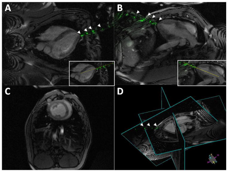
Figure 2. Real-Time MRI During LV Access.
Real-time magnetic resonance imaging (MRI)-delineated 3-dimensional trajectory for the active needle by continuously updating 2 perpendicular long-axis imaging planes for needle guidance (A,B), 1 short-axis plane for monitoring left ventricular (LV) function (C), and a 3-dimensional representation of relative positioning for these 3 planes (D). The dotted yellow line in A and B insets indicate a trajectory plan for potential aortic valve intervention. This trajectory is updated continuously as the needle is advanced toward the heart. Needle markers are evident throughout the procedure (arrowheads). Also see Online Video 1.

