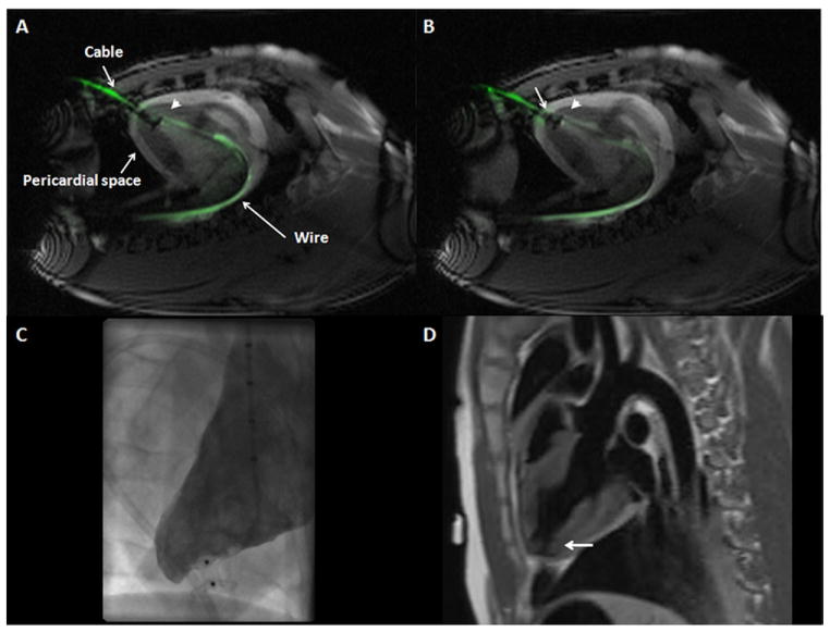
Figure 5. Muscular VSD Occluder Deployment Inside Myocardial Puncture Tract Guided by Real-Time MRI.
(A,B) “Permissive pericardial tamponade” separated the parietal from the visceral pericardium by temporary instillation of fluid into the pericardial space (“pericardial space”). (A) The “endocardial” (distal) disk (arrowhead) is released inside the LV cavity in proximity to the endocardial surface. Also visible in this image is the active delivery cable (“cable”) and back-up guidewire (“wire”). (B) The “epicardial” (proximal) disk was released against the epicardium (arrow) without entrapping the pericardium. Also see Online Video 2. (C) Early x-ray ventriculography showed occlusion of the puncture tract without extraventricular contrast leakage, and (D) MRI verified the correct position of the device inside the myocardial tract (black, indicated by the arrow) in a dark-blood acquisition. Abbreviations as in Figures 1 and 2.

